
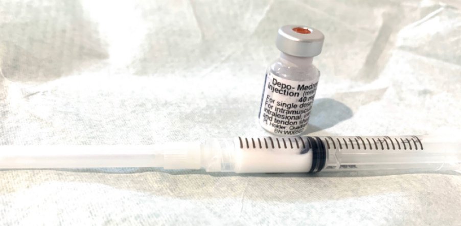
Should steroid injections for Morton’s neuroma be given without ultrasound guidance?
Table of Contents
Introduction
A 2016 study, that is frequently cited today, found minimal differences in outcomes, between Morton’s neuroma that had been injected with steroids using ultrasound guidance, compared to those injected without ultrasound guidance. This finding prompted some to argue “if the results are broadly similar why bother with ultrasound?”
So, we ask if ultrasound screening and guidance for a steroid injection for Morton’s neuroma are really necessary.
First, let us take a closer at this study
The 2016 study involved 45 feet of 36 patients. Two roughly equal groups received steroid injections from a single clinician, who was both an expert in ultrasound imaging and neuroma injections.
In the first group, the ultrasound guidance was turned on, in the second group, the machine was switched on, but the clinician only pretended to use imaging, when actually the imaging was disabled. Neither group knew when the imaging was turned on or off, so the patients were ‘blinded’, which is a good thing, as the patient’s expectations were less likely to influence the results.
The results showed that at 12 months, 48% of those that had ultrasound-guided injections were back to square one, compared to 55% that had unguided injections. In other words, a 48% failure rate for guided injections versus a 55% failure rate for non-guided injections (1), broadly speaking similar outcomes, with a modest 7% superiority for guided injections.
However, the results of this study can’t really be applied to the average person in the street with Morton’s neuroma.
This is because, unlike in real life, the study pre-screened the participants with ultrasound imaging to control any ‘confounding variables’ or unexpected reasons that could affect the study outcomes.
For example, if someone who was earmarked for the study had a pre-study screening scan that showed they had an arthritic toe joint for example, or a plantar plate tear etc. they weren’t allowed to participate in the study, as the study was only interested in people with Morton’s neuroma and nothing else. By excluding such people from the study, the researchers ensured that the main difference between the two groups being measured was the use or non-use of ultrasound guidance.
If you look at how the study was conducted, you will see that both groups of patients had, prior to the study, been assessed with ultrasound to confirm the diagnosis, and ‘weed out’ other non-neuroma conditions such as plantar plate tears, that can closely resemble Morton’s neuroma.
So, as you can see, what happened in the study is not reflective of what happens in real life. In practice, patients who receive steroid injections without ultrasound guidance rarely, if ever have ultrasound screening beforehand.
As such, inevitably there will be people that are injected ‘blindly’ for suspected Morton’s neuroma who either don’t have Morton’s neuroma, or they have other issues such as plantar plate tears, ganglion cysts etc, and as such are less likely to do well from a steroid injection. In the ‘real world’ patients having steroid injections tend to fall into one of two camps, those that have ultrasound screening and ultrasound-guided injections and those who have no ultrasound screening and have their injection blind.
Why is pre-injection ultrasound screening preferable?
Firstly, and most importantly, a scan is needed to rule out another distinct condition that can closely resemble or mimic a neuroma, a condition that might be present in up to 40% of those suffering from forefoot pain, a condition that can be made worse and not better with a steroid injection. A condition in its early stages, without ultrasound imaging, is hard to spot. The condition is known by various names including, ‘plantar plate tear’ “MTPJ instability” “pre-dislocation syndrome”, and in its most advanced stage ‘crossover toe deformity, seen below where the 2nd toe has crossed over toward the great toe.
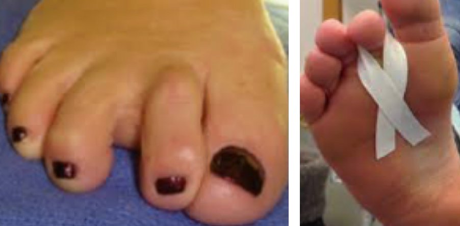
Plantar plate tears are problematic because they cause very similar symptoms to Morton’s neuroma. However, they are often treated with taping only seen above). The similarity between the two conditions can cause confusion and misdiagnosis, somewhere between 3-14% of patients with plantar plate tears end up being misdiagnosed with Morton’s neuroma, unfortunately, some end up having needless and unsuccessful neurectomy surgery (2,3,4).
Ultrasound imaging should lead to better outcomes.
If ever a future study could gain ethics approval to compare a large group of non-scanned steroid-injected suspected Morton’s neuroma sufferers with a large group of scanned injected sufferers, the study would most likely show a much bigger difference between the outcomes of the scanned and non-scanned, way in excess of the 7% advantage described in the 2016 study. This would be the case simply because there is a strong possibility that the non-scanned group would contain people with plantar plate tears that could be made worse by steroid injections.
What do the plantar plates do and why do they matter? And who gets plantar plate tears?
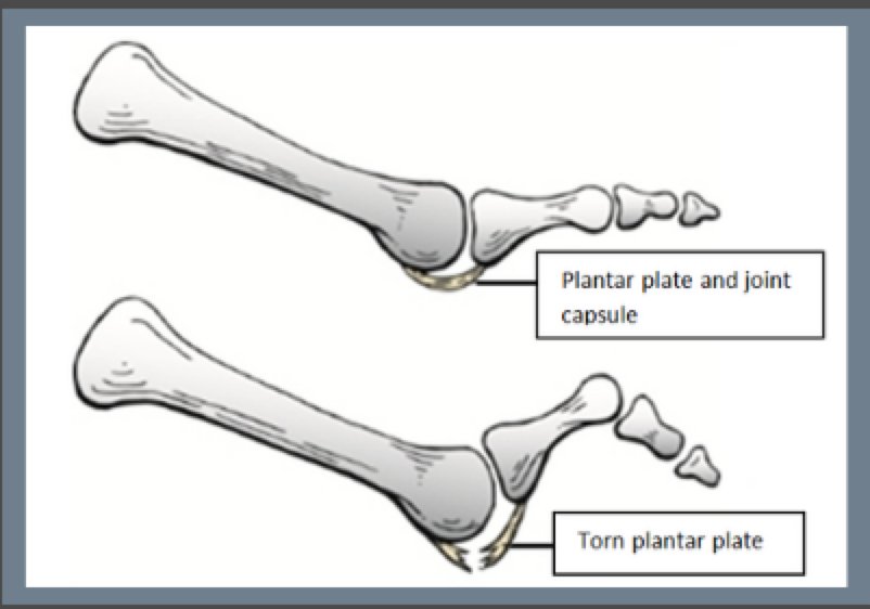
Plantar plates are ligament-like structures made from collagen, that keep our toes in contact with the ground when we walk or stand, they also help stabilise the toe joints, preventing excessive upward movement (dorsiflexion) of the toe.
Plantar plate tears mostly occur around the 2nd toe and are commonly seen in athletes who have forcefully bent their toe too far upwardly, they also start to occur as part of the ageing process. The number of people with planar plate tears in the general population is unknown, however, they are not uncommon in women in their 50s and older.
Plantar plate tears much like Morton’s neuroma seem to affect women far more commonly than men, at a ratio of 10:1. Plantar plate tears tend to cause pain, but not always. On rare occasions, we see patients with small tears, who have no pain, and yet can’t remember any past episodes of pain at the site of the tear. This suggests that some can have painless or silent tears.
How common are plantar plate tears in people with forefoot pain?
A 2014 study, that looked at the MRI scan results of 96 patients that had been referred for MRI because of forefoot pain found that 40% of patients had plantar plate tears, almost all without coexisting Morton’s neuroma (5).
What happens when a plantar plate tears?
When plantar plates tear, either wholly or partially, they can generate considerable pain and internal swelling around the metatarsal phalangeal joint, and like Morton’s neuroma the sensation of walking on a marble, as well as burning, throbbing and pins and needles. Torn plantar plates frequently enlarge, they normally measure 2-3mm, however, after a tear they fill with fluid, and can thicken up to 8 mm (6). This enlargement can impinge the nearby interdigital nerve causing neuroma-like symptoms, such as burning, pins and needles and numbness, ultimately torn plantar plates can lead to Morton’s neuroma formation because they compress the interdigital nerve.
Why are steroid injections a potential problem?
The concern with steroid injections is due to the known risk that they pose for causing rupture to soft tissue structures that contain collagen, such as the plantar plates, tendons and ligaments.
Over the decades there have been numerous case reports of such ruptures, more serious adverse events, such as rupture, occur between 1- 6% after steroid injections (7).
As such, there is a risk that a steroid injection, given near a plantar plate, that might already be degenerative can make matters worse. A steroid injection could progress a pre-existing partial incomplete tear into a complete ‘full thickness’ tear, resulting in a floating toe or crossover toe deformity. Furthermore, there is evidence from animal studies that steroid injections cause fragmentation of collagen, and delayed healing in structures comprised of collagen (8-13).
The plantar plate conundrum:
A steroid injection that may help Morton’s neuroma could make an undiagnosed plantar plate issue worse. For this reason alone, there is a strong argument that all steroid injections for Morton’s neuroma should be guided or at the very least the person receiving the injection should have been pre-screened.
When it comes to steroid injections it seems size does matter- if a neuroma is over 6 mm a steroid injection is unlikely to help.
Another compelling reason to have a scan is to determine the size of the neuroma. The size of the Morton’s neuroma prior to injection does seem to play an important role in outcomes. A healthy nerve typically measures around 2 mm in diameter, so a small neuroma is around 3-4mm in diameter, a small to medium-sized neuroma is between 4.5mm- 5.5 mm, a medium to large neuroma is between 6-8 mm and large to very large 10-14mm.
Understanding the size of your neuroma (in diameter) is important because neuromas greater than 5 mm in diameter rarely get more than short to medium-term relief from steroid injections (13). Furthermore, a large study of 201 Morton’s neuroma sufferers found that the threshold for steroid injection failure was 6.3 mm, the study demonstrated “… neuromas with a size larger than 6.3 mm can be suspected to require further treatment with a certainty of 84%.” (15).
Are there any alternatives to steroid injections and conventional surgery that can be curative even for very large Morton’s neuroma?
A cryosurgery study involving 24 large neuromas (average size 12.7mm) showed at 19.7 months post-treatment that 80% had a high satisfaction score with no incidence of stump neuroma formation (16). Cryosurgery has now been performed for Morton’s neuroma since 1996 and has proved to be a safe and effective form of treatment for Morton’s neuroma, furthermore, cryosurgery does not rely on the use of steroid injections.
References
- Mahadevan D, Attwal M, Bhatt R, Bhatia M. Corticosteroid injection for Morton’s neuroma with or without ultrasound guidance: a randomised controlled trial. Bone Joint J. 2016 Apr;98-B(4):498-503. doi: 10.1302/0301-620X.98B4.36880. PMID: 27037432.
- Coughlin MJ. Crossover second toe deformity. Foot Ankle. 1987;8:29-39.
- Coughlin MJ. Second metatarsophalangeal joint instability in the athlete. Foot Ankle. 1993;14:309-319.
- Kaz AJ, Coughlin MJ. Crossover second toe: demographics, etiology, and radiographic assessment. Foot Ankle Int. 2007;28:1223-1237.
- Umans H, Srinivasan R, Elsinger E, Wilde GE. MRI of lesser metatarsophalangeal joint plantar plate tears and associated adjacent interspace lesions. Skeletal Radiol. 2014 Oct;43(10):1361-8. doi: 10.1007/s00256-014-1920-8. Epub 2014 Jun 1. PMID: 24880715
- Blitz NM, Ford LA, Christensen JC. Plantar plate repair of the second metatarsophalangeal joint: technique and tips. J Foot Ankle Surg. 2004; 43(4):266-70.
- Brinks A, Koes BW, Volkers AC, Verhaar JA, Bierma-Zeinstra SM. Adverse effects of extra-articular corticosteroid injections: a systematic review. BMC Musculoskelet Disord. 2010 Sep 13;11:206. doi: 10.1186/1471-2474-11-206. PMID: 20836867; PMCID: PMC2945953.
- Kleinman M, Gross AE. Achilles tendon rupture following steroid injection. Report of three cases. J Bone Joint Surg Am. 1983;65:1345–7. [PubMed] [Google Scholar]
- Stannard JP, Bucknell AL. Rupture of the triceps tendon associated with steroid injections. Am J Sports Med. 1993;21:482–5. [PubMed] [Google Scholar]
- Balasubramaniam P, Prathap K. The effect of injection of hydrocortisone into rabbit calcaneal tendons. J Bone Joint Surg Br. 1972;54:729–34. [PubMed] [Google Scholar]
- Kapetanos G. The effect of the local corticosteroids on the healing and biomechanical properties of the partially injured tendon. Clin Orthop Relat Res. 1982;163:170–9. [PubMed] [Google Scholar]
- Oxlund H. The influence of a local injection of cortisol on the mechanical properties of tendons and ligaments and the indirect effect on skin. Acta Orthop Scand. 1980;51:231–8. [PubMed] [Google Scholar]
- Wiggins ME, Fadale PD, Barrach H, Ehrlich MG, Walsh WR. Healing characteristics of a type I collagenous structure treated with corticosteroids. Am J Sports Med. 1994;22:279–88. [PubMed] [Google Scholar]
- Makki D, Haddad BZ, Mahmood Z, Shahid MS, Pathak S, Garnham I. Efficacy of corticosteroid injection versus size of plantar interdigital neuroma. Foot Ankle Int. 2012 Sep;33(9):722-6. doi: 10.3113/FAI.2012.0722. PMID: 22995258.
- Park, Young Hwan; Lee, Ja Woon; Choi, Gi Won; Kim, Hak Jun (2017). Risk factors and the associated cutoff values for failure of corticosteroid injection in treatment of Morton’s neuroma. International Orthopaedics, (), –. doi:10.1007/s00264-017-3707-8
- Cazzato RL, Garnon J, Ramamurthy N, Tsoumakidou G, Caudrelier J, Thenint MA, Rao P, Koch G, Gangi A. Percutaneous MR-Guided Cryoablation of Morton’s Neuroma: Rationale and Technical Details After the First 20 Patients. Cardiovasc Intervent Radiol. 2016 Oct;39(10):1491-8. doi: 10.1007/s00270-016-1365-7. Epub 2016 May 17. PMID: 27189181.

Mr Weaver has a special clinical interest in podiatric regenerative medicine and was among the first clinicians in Europe—and the first in the UK—to offer cryosurgery (cryoablation) for Morton’s neuroma. Driven by a commitment to excellence in patient care, he focuses on delivering precise, evidence-based treatments that prioritise long-term recovery and patient comfort.
Over the past 18 years, he has performed approximately 20,000 procedures for Morton’s neuroma and bursitis. This extensive experience allows him to provide a highly personalised approach, ensuring every patient receives the specialist attention required for the best possible outcomes.
His professional memberships include the Institute of Podiatrists, the International Society of Cryosurgery, and the British Medical Ultrasound Society.