

Case Study: Cryosurgery for Stump Neuroma
Table of Contents
Introduction:
The following is a case study of one of our stump neuroma success stories. Stump neuromas can cause life-limiting discomfort and loss of enjoyment. Stump neuroma occurs in at least 30% of those that have undergone excision surgery. They are caused by a proliferation of fibrous scar tissue around the remaining elements of the nerve.
Stump Neuroma is generally treated with steroid injections, which can definitely help, or with further surgery, which has a very high rate of stump neuroma reoccurrence. Many who have Stump Neuroma do not realise that there is a safe treatment that can, as in the case of our subject, restore normal pain-free life. If you have a stump neuroma, don’t wait 13 years. Get in touch with The Barn Clinic today.

Case Study Overview:
A 59-year-old female patient with a 13-year history of stump neuroma pain who was treated with Cryosurgery at The Barn Clinic, London in September 2020 and was pain-free at 6 months follow up.
Patient description:
The following is a case study of a 59 year-old female district nurse. A non-smoker, who enjoys occasional alcohol consumption and isn’t overweight. Taking no regular medications, and without any allergies.
Case history:
The patient developed intermittent pain in her right foot in the 1980’s, which progressed over the years to become a constant discomfort. The patient attributed the onset of her symptoms to working long shifts as a nurse that involved a lot of heavy manual lifting and handling of patients. In 2006, the patient was assessed by a specialist foot & ankle clinic and diagnosed with Morton’s neuroma in the right 2nd/3rd interspace.
X-Rays taken at that time were essentially normal and ruled out any other obvious bony cause of her pain. The patient subsequently received physiotherapy, foot orthotics (with and without metatarsal domes or bars) and steroid injections (with and without ultrasound guidance) to the right 2nd/3rd interspace. Unfortunately, none of these treatments gave any improvement to the patient. In 2007, the patient underwent dorsal (from above) excision of a well-defined Morton’s neuroma.
Due to persisting pain and return of the neuroma the area was re-operated on in 2008, this time from below and by another surgeon and then, again by a third surgeon in 2010. Unfortunately, none of these operations gave the patient any improvement, and if anything made matters worse.
In 2014, the patient had gone on to develop pain beneath left 3rd-4th toes and was re-referred to the foot & ankle clinic who diagnosed a Morton’s neuroma in the left 3rd-4th interspace, from a physical exam. They also arranged an MRI scan of both feet that revealed the present of a bursa (sac of fluid) and scar tissue in the right 2nd/3rd interspace, and a bursa and Morton’s neuroma in the left 3rd/4th interspace.
At that stage, the patient was offered further surgery to try and improve the scar tissue, and excision of the neuroma in the left foot. Having seen no improvement from her previous surgeries the patient decided to decline the offer of surgery and was subsequently discharged as a patient.
The patient managed her ongoing symptoms by avoidance of prolonged standing and walking.
Past Imaging:
To date, imaging had included X-Rays prior to her original excision surgery in 2007, and an MRI in 2014, showing a lot of scar tissue in the right forefoot.
The patient’s description of her pain and disability:
Upon presentation to myself in September 2020, the patient described constant right forefoot pain localised to the underside of the forefoot in the region beneath the right 2nd, 3rd and 4th toes. In addition to the pain in the bottom of her foot, the patient also described hypersensitivity on the upper aspect of the foot, localised to the scar of the original surgery. The hypersensitivity meant that even very light touch or pressure caused discomfort. When asked to describe the nature of her pain the patient said her foot “experienced burning, electrical and shooting sensations, that were sometimes sharp and also dull in nature, with pins and needles and numbness along with the sensation of having a stone-in-the-shoe”.
When asked what worsened her symptoms the patient replied, “Standing, massage and heat”. When asked what helps the symptoms the patient replied, ”Cold and sometimes, oddly, walking. However, walking makes the pain of my left neuroma worse. When asked what things doesn’t seem to make a difference, the patient replied “sitting and taking pain killers”.
When I asked the patient to grade the severity of her pain the patient said that “as a nurse, over the years I have learnt how do deal with pain”. Accordingly, she graded her constant pain as 3/10 (10 being the worst pain imaginable) but when the pain hits it’s peak, she graded the pain in her right foot as 6.5/10, and the left foot as 2.5/10 rising to 5/10 at its worst.
On Physical examination:
I noted her feet were moderately pronated but otherwise well aligned. There was some arthritis of the left big toe joint that was stiffened with a reduced range of motion.
When squeezing the metatarsal bones together and pushing up with my thumb I felt a distinct clicking sensation in the right 2nd/3rd interspace and the left 3rd/4th interspace, indicating the presence of abnormal tissue in the right 2nd/3rd interspace and a Morton’s neuroma in the left 3rd/4th.
Ultrasound imaging:
Revealed an 8mm wide region of dense, fibrous scar tissue surrounding the bulbous down turned stumps of the remaining nerve that had received the three operations in the right foot. In addition, I noted a bursa or sac of fluid sitting above and around the nerve and medium sized sac of fluid in the right 3rd/4th interspace, and degeneration of one of the main tendons in the right ankle called the tibialis posterior tendon. I also found a 6.9mm wide Morton’s Neuroma in the left 3rd/4th interspace.
Treatment plan:
- Cryosurgery to address to right 2nd stump neuroma
- Cryosurgery to address the bursa sitting above the right 2nd/3rd stump neuroma
- Cryosurgery to the left 3rd/4th Morton’s neuroma.
- Conservative management options of the right tibialis posterior tendon.
The expected outcome of the proposed treatment:
Stump neuroma: Due to the extent of past surgery to the right 3rd/4th nerve, there was a wide rage of possible outcomes that ranged from:
- No improvement
- Modest improvement
- Substantial improvement
- Total resolution of pain
Morton’s Neuroma: The expected outcome for the left 3rd/4th neuroma was much more positive, in that, I expected either total resolution or substantial improvement.
Actual outcome following the proposed treatment – 28th March 2021:
Morton’s Neuroma: The patient reported the cryosurgery to the left 3rd/4th interspace had been totally successful reporting 0/10 pain during normal activities, and no pain after recreational activity (usually a 5 mile walk in the countryside).
Stump Neuroma: The patient again reported an identical outcome to left foot in that she no longer felt any pain at the site of the right 2nd/3rd stump. The patient reported that she had previously been limited to only being able to walk 10 minutes before being forced to stop. Post cryosurgery the patient reports being able to walk for 5 miles without any discomfort.
Clinical findings at 24 Week Follow-Up:
Right foot:
The patient was negative for clicking and pain on the squeeze and pinch test to the right 2nd/3rd interspace, however, the patient was tender in the right 3rd/4th interspace during the pinch test. Ultrasound imaging demonstrated a substantial reduction in the right 2nd/3rd inter-metatarsal stump neuroma scar tissue. The appearance of the tissue compared favourably to that of a healthy interspace. Ultrasound imaging also demonstrated the bursal tissue sitting above the stump neuroma had also disappeared since her cryosurgery.
Ultrasound imaging to the right 3rd/4th interspace (the area that was slightly tender during the pinch that had no previous issues or treatment) demonstrated a small amount of fluid around the nerve. When questioned, the patient stated that she is only aware of any discomfort in the right 3rd/4th region of her forefoot if she walks over 5 miles, and as she rarely exceeds 5 miles this is currently not an issue to her that needs treatment.
Left foot:
The interspaces in the left forefoot were negative for clicking or pain during the squeeze and pinch tests. The 6.9mm Morton’s neuroma identified with previous imaging was no longer present.
Discussion
Pain and disability stemming from stump neuroma can be very difficult to treat. Further surgery frequently results in additional scar tissue formation. Steroid injections can sometimes be helpful in remodelling scar tissue, however, repeated steroid injections can cause ligament injury to the nearby plantar plate ligaments and subsequent painful toe deformities. In addition to the risk of plantar plate tear, two or more steroid injections can cause loss of protective fat tissue in the forefoot, making the metatarsal heads feel more prominent and uncomfortable during walking.
This case study describes how cryosurgery can offer an effective treatment alternative to further surgery and steroid injections.
Cryosurgery delivered via a probe rather than a spray has been shown to be an effective and safe way of reversing scar tissue, especially keloid scars (1). Cryosurgery shrinks back scar tissue depriving the scar tissue of its blood and oxygen supply (2) causing the abnormal tissue to be broken down and reabsorbed.
Cryosurgery has been used over the last 30 years to treat hypertrophic (overgrown) and keloid scars with a high degree of safely and success (3,4,5,6,7,8,9,10,11,12,13,14).
References
- Goldenberg G, Luber AJ. Use of intralesional cryosurgery as an innovative therapy for keloid scars and a review of current treatments. J Clin Aesthet Dermatol. 2013 Jul;6(7):23-6. PMID: 23882311; PMCID: PMC3718752.
- Har-Shai Y, Mettanes I, Zilberstein Y, et al. Keloid histopathology after intralesional cryosurgery treatment. J Eur Acad Dermatol Venereol. 2011;25(9):1027–1036.
- Thomas DW, Hopkinson I, Harding KG, Shepherd JP. The pathogenesis of hypertrophic/keloid scarring. Int J Oral Maxillofac Surg. 1994;23(4):232–236. Berman B, Zell D, Romagosa R. Keloid scarring. In: Lebwohl MG, Heymann WR, Berth-Jones J, Coulson I, eds. Treatment of Skin Disease Comprehensive Therapeutic Strategies. 2nd ed. Philadelphia, PA: Elsevier Limited; 2006:314–317.
- Berman B, Zell D, Romagosa R. Keloid scarring. In: Lebwohl MG, Heymann WR, Berth-Jones J, Coulson I, eds. Treatment of Skin Disease Comprehensive Therapeutic Strategies. 2nd ed. Philadelphia, PA: Elsevier Limited; 2006:314–317.
- Mende B. Treatment of keloids by cryotherapy. Z. Hautkr. 1987;62(18):1348,1351–1352,1355.
- Zouboulis CC, Blume U, Büttner P, Orfanos CE. Outcomes of cryosurgery in keloids and hypertrophic scars: a prospective consecutive trial of case series. Arch Dermatol. 1993;129(9):1146–1151.
- 7. Rusciani L, Paradisi A, Alfano C, et al. Cryotherapy in the treatment of keloids. J Drugs Dermatol. 2006;5(7):591–595.
- Rusciani L, Rossi G, Bono R. Use of cryotherapy in the treatment of keloids. J Dermatol Surg Oncol. 1993;19(6): 529–534.
- Har-Shai Y, Amar M, Sabo E. Intralesional cryotherapy for enhancing the involution of hypertrophic scars and keloids. Plast Reconstr Surg. 2003;111(6):1841–1852. 15.
- Lawrence WT. In search of the optimal treatment of keloids: report of a series and a review of the literature. Ann Plast Surg. 1991;27(2):164–178.
- Weshahy AH. Intralesional cryosurgery. A new technique 16. using cryoneedles. J Dermatol Surg Oncol. 1993;19(2): 123–126.
- Gupta S, Kumar B. Intralesional cryosurgery using lumbar puncture and/or hypodermic needles for large, bulky, 17. recalcitrant keloids. Int J Dermatol. 2001;40(5):349–353.
- Har-Shai Y, Dujovny E, Rohde E, Zouboulis CC. Effect of skin surface temperature on skin pigmentation during contact and intralesional cryosurgery of keloids. J Eur Acad Dermatol Venereol. 2007;21(2):191–198.
- Har-Shai Y, Brown W, Pallua N, Zouboulis CC. Intralesional cryosurgery for the treatment of hypertrophic scarsand keloids. Plast Reconstr Surg. 2010;126(5):1798–1800.
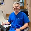
Mr Weaver has a special clinical interest in podiatric regenerative medicine and was among the first clinicians in Europe—and the first in the UK—to offer cryosurgery (cryoablation) for Morton’s neuroma. Driven by a commitment to excellence in patient care, he focuses on delivering precise, evidence-based treatments that prioritise long-term recovery and patient comfort.
Over the past 18 years, he has performed approximately 20,000 procedures for Morton’s neuroma and bursitis. This extensive experience allows him to provide a highly personalised approach, ensuring every patient receives the specialist attention required for the best possible outcomes.
His professional memberships include the Institute of Podiatrists, the International Society of Cryosurgery, and the British Medical Ultrasound Society.