
Intermetatarsal bursitis is one of the most common causes of pain in the ball of the foot — and one of the most common reasons a Morton’s neuroma is misdiagnosed. At The Barn Clinic we treat both, very often together, with minimally invasive cryosurgery.
What Is a Bursa, and Why Does It Become a Problem?
Bursae are small sacs of fluid that act as buffers, cushioning the soft tissues of the forefoot against the pressure they take with every step. When a bursa between the metatarsals becomes irritated, it can turn inflamed, enlarged and fibrotic — and that is intermetatarsal bursitis.
A common cause of forefoot pain. As the bursa swells, it takes up space in an already crowded part of the foot and presses on the small nerve that runs alongside it.
Often mistaken for a Morton’s neuroma. That pressure on the nerve produces burning, tingling and the classic feeling of “walking on a pebble” — the very same symptoms as a Morton’s neuroma. On symptoms alone, the two are almost impossible to tell apart.
Intermetatarsal bursitis · part 1
Intermetatarsal Bursitis and Morton's Neuroma: Telling Them Apart
What the intermetatarsal bursa does, how chronic bursitis can lead to a Morton's neuroma, and why the two are so often confused.
- What the intermetatarsal bursa does. Bursas are small sacs of synovial fluid sitting around joints and between the structures of the forefoot that act as lubricating mechanical buffers.
- Bursitis as a precursor to Morton's neuroma. When a forefoot bursa becomes chronically inflamed, fibrotic and enlarged, it presses on the adjacent plantar nerve — causing nerve compression, thickening and fibrosis that leads to a Morton's neuroma.
- High rate of co-existence and misdiagnosis. Around 40% to 50% of patients presenting with Morton's neuroma have co-existing bursopathy. Imaging studies show that only around 20% of patients who think they have a neuroma actually have one on scan — the remaining 80% have other causes, most frequently intermetatarsal bursitis.
- Why clinical separation is difficult. Because an inflamed bursa compressing the nerve generates identical symptoms to a Morton's neuroma, diagnostic ultrasound imaging is essential to separate the two.
Read the full transcript
Hi, I'm Robin. I'm on holiday, I'm here with my lovely wife Clare, and before I have my first beer, I'm going to talk to you about intermetatarsal bursitis. Bursas: what are they, what do they do, why do they become a problem, and more importantly, what can you do to fix them?
So bursas are little sacs of fluid that you find, typically around joints, and they are buffers. Bursas contain synovial fluid, which keeps everything lubricated. Commonly you find bursas in the knee, in the hip, and in the elbow — they're very common places to find bursas — and you also get them in the forefoot, because you've got a lot of structures in the forefoot that have the potential to rub against each other. So bursas form a buffering function; that's what they do.
Can they become a problem in themselves? Yes, they can, because the bursas themselves can actually become inflamed, fibrotic, and enlarged. And when they become very enlarged, inflamed, and fibrotic, they press on the nerve. When they press on the nerve, that causes compression, which causes the nerve in turn to change: the nerve thickens up, becomes fibrotic itself, and then you have a neuroma. So many people think that bursitis is actually a precursor to Morton's neuroma, and that does make sense.
In terms of what I see in my day-to-day practice in the clinic, the majority of patients who present with Morton's neuroma actually do have co-existing bursopathy. It's estimated that about 40% to 50% of patients with Morton's neuroma have co-existing bursopathy.
There are various studies that bear that out about bursopathy and its frequency. Most patients that think they've got a neuroma actually have bursitis. Only about 20% of patients that think they have a neuroma, when they get investigated with imaging, actually have a neuroma. The other 80-odd percent of patients have other causes, and one of the most frequent causes is bursopathy, or bursitis.
Morton's neuroma and bursopathy are very hard to distinguish clinically because they both generate the same symptoms. That inflamed sac of fluid bears down on the nerve and compresses it. And if the nerve is being compressed, that actually generates identical symptoms to Morton's neuroma — it mimics Morton's neuroma. So clinically, it can be hard to separate the two.
The Neuroma–Bursa Complex
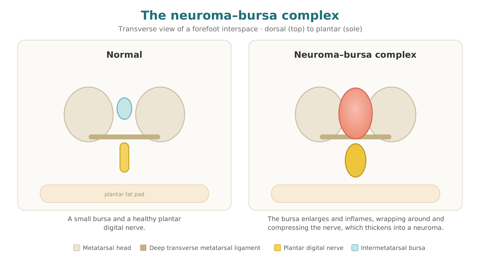
In many patients, the bursa and the nerve are really one problem. As the bursa thickens, it can wrap around and compress the nerve — and that ongoing compression is thought to be one of the ways a Morton’s neuroma develops in the first place.
When Mr Weaver treats a Morton’s neuroma with cryosurgery, he frequently finds the nerve is being strangled by an enlarged bursa that has grown around it. This is why both have to be treated together: address the nerve but leave the bursa, and the result is far less reliable. In our experience a large share of patients who are told they have a neuroma in fact have this neuroma–bursa complex.
Why an Accurate Diagnosis Comes First
Not everyone who is told they have a Morton’s neuroma actually has one. When forefoot pain is investigated with imaging, only a proportion of patients turn out to have an isolated neuroma; in many, the real culprit is bursitis, or the two combined.
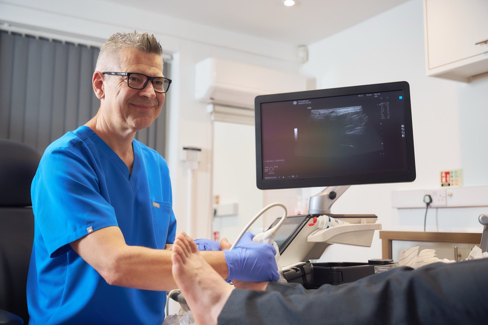
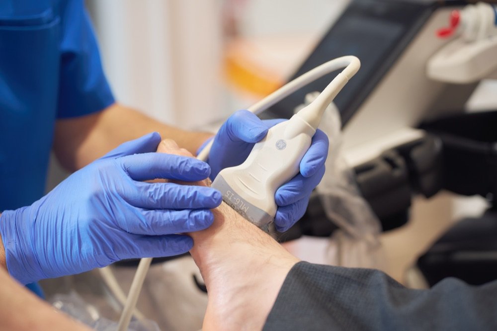
That is why every assessment at The Barn Clinic includes dynamic, high-resolution ultrasound, carried out in the clinic and in real time. It lets us look at the bursa and the nerve separately, find the true source of the pain, and aim treatment at the right structure. The published studies behind our approach are available on request.
How Is Intermetatarsal Bursitis Treated?
Start with the cause. If footwear is the problem — shoes that are too narrow, or a heel that tips weight onto the ball of the foot — that is the first thing to change. Many patients are helped by a wider toe box and flatter, “zero-drop” shoes. Where the foot’s mechanics play a part, bespoke orthotics can offload the painful area.
Be cautious with steroid injections. A cortisone injection can calm the inflammation for a while, and it has its place. But injected into the ball of the foot it can waste away the protective fat pad — some patients lose up to 20% of that cushioning from a single injection, and it does not grow back. Losing the padding that shields the forefoot can leave you worse off in the long run.
Intermetatarsal bursitis · part 2
Footwear, Orthotics, and the Risks of Steroid Injections
The mechanical changes that take pressure off the forefoot, where orthotics help, and the long-term drawback of corticosteroid injections.
- Addressing mechanical overload via footwear. Narrow shoes and elevated heels throw weight forward onto the ball of the foot. Raising the heel reduces forefoot contact area and shifts load medially beneath the first metatarsal head, increasing pressure there even though total forefoot load is unchanged (Corrigan et al., Foot Ankle 1993). At dress-heel heights the effect is large: going from a 2 cm to a 6 cm heel raised peak pressure under the central metatarsals by 30%, and the pressure-time integral by 48% (Speksnijder et al., The Foot 2005, ten women). Conventional trainers sit far lower, at roughly an 8 to 12 mm heel-to-toe drop, but the direction of effect is the same; zero-drop shoes, where the heel sits level with the ball of the foot, remove it.
- Correcting biomechanics with orthotics. Both excessive pronation (feet turning in) and supination (high arches) predispose patients to bursitis and can be addressed with orthotics inside the shoes.
- The drawbacks of steroid injections. While corticosteroid injections offer an immediate anti-inflammatory effect, a single steroid injection can cause patients to lose up to 20% of their protective plantar fat pad volume — a loss that does not grow back in the ball of the foot and compounds mechanical pain.
Read the full transcript
First of all, I say to my patients, if you've got bursitis, we need to think about what might be causing it in the first place. So, if there's an obvious candidate — if your shoes are just too small, too narrow, or if you've got a high heel that's throwing all your weight forward onto the ball of your foot — I'd say just address those issues first of all.
And then, if your bursitis is related more to activity, maybe think about appropriate footwear for that activity. Commonly, zero-drop shoes are used. Zero-drop shoes, if you're not familiar with them, are trainers where the heel sits at the same level as the ball of the foot.
If that doesn't work, or it's too far gone, I think the next step along would be orthotics. If you've got a mechanical imbalance in your foot — if your feet are pronated and turn excessively in, or if, on the other hand, you've got very high arches — both being pronated and supinated is a predisposing factor for the development of bursitis. So if you've got any of those factors in play, deal with those, typically with orthotics that you have inside your shoes.
If you've tried all of that and you're still suffering with bursitis, then what do you do? The NHS typically would offer you steroid injections into and around the bursa. Steroid injections can be very helpful for certain patients; they can offer an immediate anti-inflammatory effect.
However, a common problem that I see with steroid injections is that they can be associated with loss of fat tissue at the site of the injection. If you lose your protective fat tissue in the ball of the foot, it just compounds the problem. Unfortunately, the ball of the foot is the one place that you don't get fat tissue back. It's an irony that as we get older, we tend to develop fat tissue around our abdomen and all over the place, but the one place where you need some fat tissue is the one place where you lose it as you get older. Some people can lose up to 20% of their protective fat tissue volume from a single steroid injection. For that reason, I'm not very keen on steroid injections — but there is a place for them.
Cryosurgery for Bursitis and the Neuroma–Bursa Complex
When the simple measures have not worked, cryosurgery offers a minimally invasive alternative to open surgery.
Performed in clinic, under local anaesthetic. It is a day-case procedure — no hospital stay, no general anaesthetic, no stitches.
Treats the whole complex at once. Guided by ultrasound, a fine probe is placed at the affected interspace and a controlled freeze treats the nerve and the surrounding bursa together.
No risk of stump neuroma. Unlike surgery to cut the nerve out, cryosurgery is nerve-sparing, so it avoids permanent numbness and the painful stump neuroma that can follow excision.
Safe and effective. Mr Weaver has performed ultrasound-guided cryosurgery since 2009, with complications being very uncommon. When patients return for follow-up imaging around six months later, we typically see the bursa settled back to normal and the nerve restored to a healthy appearance.
Intermetatarsal bursitis · part 3
Ultrasound-Guided Cryosurgery as a Restorative Treatment
How cryosurgery treats the thickened nerve and the inflamed bursa together, and what the follow-up imaging shows.
- Simultaneous resolution of neuroma and bursitis. Under ultrasound guidance the bursa is frequently seen enveloping and strangling the nerve. Addressing both the Morton's neuroma and the bursitis simultaneously with cryosurgery clears up both conditions at six-month follow-up imaging.
- A safety track record since 2009. Performed in clinic since 2009, ultrasound-guided cryosurgery for Morton's neuroma and bursopathy has a strong safety profile, with complications very few and far between.
- Restorative rather than destructive. Unlike excision surgery, alcohol injections or radiofrequency — which cause permanent, irreversible changes to the nerve on imaging — cryosurgery is restorative and rejuvenative, with both the nerve and the bursa reverting towards a normal healthy appearance on follow-up scans without negative scarring.
Read the full transcript
When it comes to Morton's neuroma and bursopathy — because the two do go hand in hand — and the treatments thereof, I think the most successful treatment by a long, long way is cryosurgery. And that's borne out by the imaging that we do with patients when we see them back for follow-up.
When we see cryosurgery patients in clinic six months down the line and we look at them on ultrasound, not only do we see, thankfully, that they've got no neuroma symptoms and no presence of the neuroma, but we also see that the bursa has cleared up. So the whole environment in the forefoot that used to contain the neuroma and the bursitis is a completely different situation when we revisit six months later. The bursopathy has gone, as has the neuroma, and patients are back again doing the kind of things that they used to do before.
When we treat a Morton's neuroma with cryosurgery, we're guiding a cryoprobe onto the neuroma, and we frequently see that the neuroma is strangled by the bursa — the bursa has actually enveloped the nerve. So by virtue of treating one, we treat the other. When we treat the Morton's neuroma, we have to address the bursitis with the cryosurgery; otherwise, the treatment doesn't work anywhere near as well.
I actually think that cryosurgery for Morton's neuroma and bursopathy is incredibly safe. I've been doing it now since about 2009, and thankfully, complications are very few and far between. Cryosurgery for bursopathy seems extremely effective. When we revisit those patients and look at them on imaging, we find typically that the whole of the area has improved. The bursa frequently has just gone back to normal — it contains a normal amount of fluid — and that pre-existing inflammation and the whole 'angry' state of the bursa has completely changed and reverted back to a normal appearance. So I think it's almost unique.
Cryosurgery for Morton's neuroma has an incredible feature that not many people actually think about or are aware of. The most striking feature, I think, of cryosurgery is that it reverts the nerve back to normal. I can't think of any other treatments that revert the nerve back to normal; it's restorative and it's rejuvenative. If you look at patients that have had cryosurgery and have had a positive outcome, you see a normal-looking, healthy nerve on imaging. I can't think of any other treatment modality that actually facilitates that.
All the other treatments for Morton's neuroma that I can think of — surgery, alcohol injections, radiofrequency, et cetera — all cause permanent, irreversible change to the nerve that can be detected on imaging. Whereas after cryosurgery, you see a restoration of what would be there normally, and it doesn't seem to cause any negative scarring or side effects. So I've found it to be a very helpful, reliable, safe, effective treatment over the years.
How Does Cryosurgery Work?
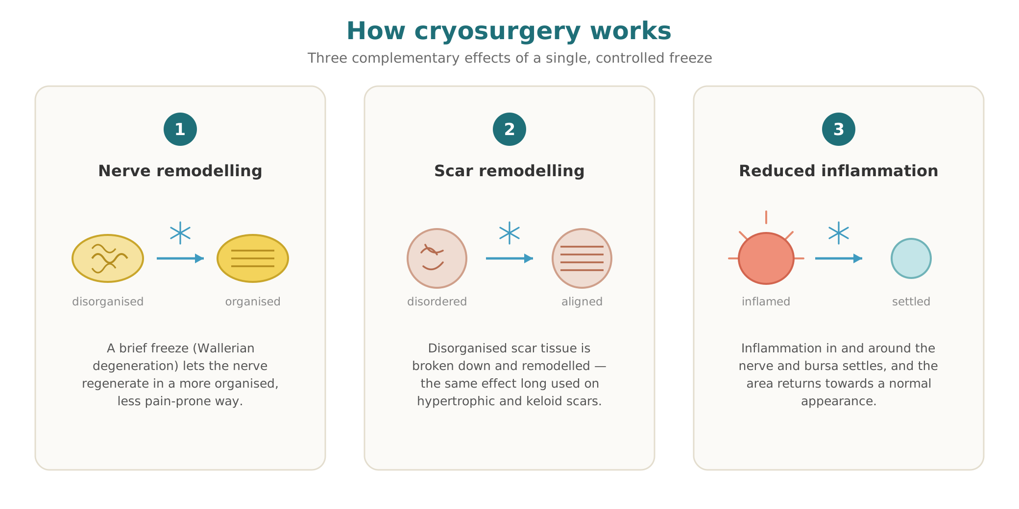
Cryosurgery seems to work in three ways at once. It remodels the nerve — a brief, controlled freeze prompts a natural process called Wallerian degeneration, after which the nerve tends to regenerate in a calmer, less painful form. It remodels scar tissue — cryosurgery has long been used to treat hypertrophic and keloid scars, and a similar effect helps break down the fibrous tissue of a chronic neuroma–bursa complex. And it calms the inflammation in and around the nerve and bursa, which is why the whole area settles.
“After 6 weeks I am cycling 100 miles a week, and playing 3 rounds of golf each week.”
Contact Us for More Information
If you have ongoing pain in the ball of the foot — or you have been told it is a Morton’s neuroma but nothing has helped — the first step is to find out what is really there. Our clinics are in Harley Street, London, and in Sheffield. Contact us or request an information pack, and we will be glad to help.